News
Rädler Group: new review on cell migration
22 Apr 2024
'Mesenchymal cell migration on one-dimensional micropatterns'
22 Apr 2024
'Mesenchymal cell migration on one-dimensional micropatterns'
We are excited to see our review article on 1D cell migration published in Frontiers in Cell and Developmental Biology. Our understanding of cell migration has immensely profited from studying cells move in one-dimensional confinements, such as on microfabricated lanes. In our article we review 1D migration assays, reflect on the advantages of such platforms and discuss popular biophysical models of cell migration. We highlight the unique opportunity of reproducible and standardised 1D assays to validate theory based on statistical measures from large data of trajectories and discuss the potential of experimental settings embedding controlled perturbations to probe response in migratory behaviour.
Johannes C. J. Heyn, Joachim O. Rädler, Martin Falcke
Mesenchymal cell migration on one-dimensional micropatterns
Front. Cell Dev. Biol., Sec. Cell Adhesion and Migration, Volume 12, 2024
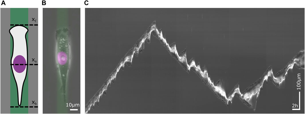
Single cell migration on a 1D micropattern. 1D micropatterns facilitate the study of mesenchymal cell migration by enabling the acquisition of large statistics. (A) Schematic sketch of a cell on a lane that has been functionalized with an extracellular matrix (ECM) protein. The migration of the cell is defined by the position of its front xf, its nucleus xn and its back xb over time. (B) A human breast cancer cell (MDA-MB-231) on a fibronectin (FN) lane. The phase contrast image visualizes the contour of the cell. The nucleus has been stained violet and the ECM protein green. Scale bar 10 µm. (C) Kymograph of a migrating cell whose trajectory displays changes in velocity and direction as well as in cell length. Time runs from left to right. The vertical axis represents the position along the center of a micropatterned lane. Horizontal scale bar 1h, vertical scale bar 100 µm.